The X-ray tube is another extremely important type of vacuum tube that still has many applications these days. Since Wilhelm Conrad Roentgen first discovered the X-ray, in 1895, they have been utilized in several fields that benefit society.

However, it is important to note that their applications are still expanding. Consequently, despite it being more than one hundred years old, X-ray tubes are going to play an irreplaceable role in many areas including medical and security scanners.
Working principle
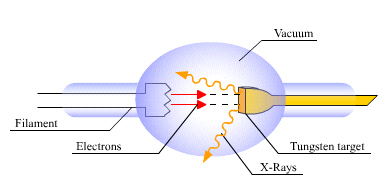
As illustrated in the diagram to the right, when the filament is heated to several thousand degrees, a large amount of electrons are released from the filament that accelerate towards the anode, which is usually made of tungsten. After the acceleration due to the potential difference of the filament (the cathode) and the target (the anode), the electrons will hit the other atomic particles of the tungsten target and rapidly decelerate. This effect produces Bremsstrahlung radiation, also known as ‘deceleration radiation’; X-rays are generated from the anode. Of course, the anode and the cathode are enclosed in a vacuum tube to prevent the filament from burning up and to prevent arcing between the cathode and anode.
Medical Application – CT scanners
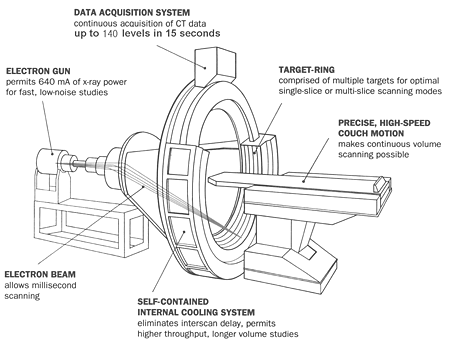
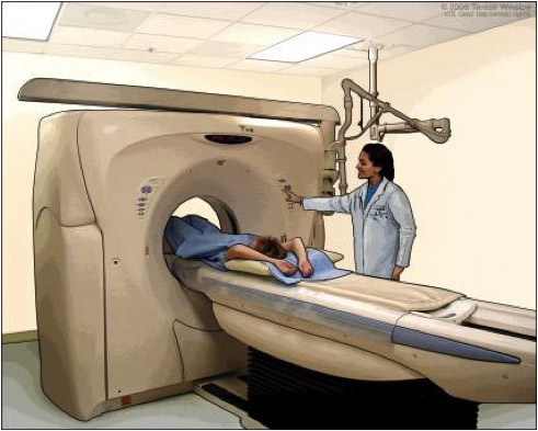
The X-ray tube is employed in the computed tomography (CT) scanner, which is an important medical device able to produce a large amount of data through a process known as “windowing”. This process is able to display various bodily structures based on their ability to block the incoming X-ray beams. After that it can reconstruct images mathematically from this data and display them in a digital form. CT was brought into clinical practice in 1972 and has been being used in almost all hospitals around the world.
Further Reading/ References:
1. http://en.wikipedia.org/wiki/X-ray_computed_tomography
2. http://www.radiologyinfo.org/en/info.cfm?PG=bodyct#part_nine

